TERUMO MEDICAL PRODUCTS Science & Industry Copy
TERUMO Medical Products
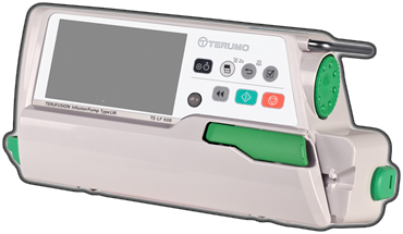
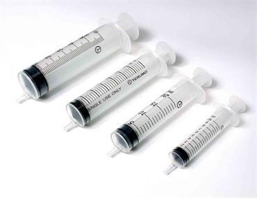
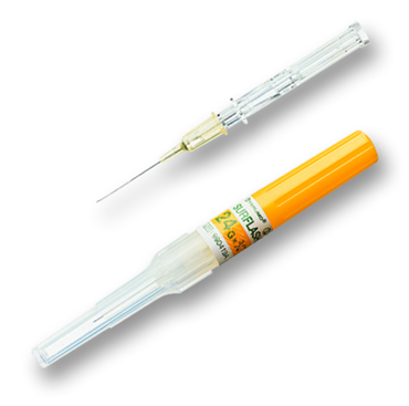
Terumo Medical Product’s main areas of expertise are injection and infusion.


ΔΙΕΓΧΕΙΡΗΤΙΚΗ ΚΥΤΤΑΡΟΜΕΤΡΙΑ ΡΟΗΣ ΣΕ ΟΓΚΟΥΣ ΕΓΚΕΦΑΛΟΥ:
ΕΞΑΤΟΜΙΚΕΥΜΕΝΗ ΧΕΙΡΟΥΡΓΙΚΗ ΕΠΕΜΒΑΣΗ
ΑΚΡΩΝΥΜΙΟ: ΔΙΕΓΧΕΙΡΗΤΙΚΗ ΚΥΤΤΑΡΟΜΕΤΡΙΑ ΡΟΗΣ
ΚΩΔΙΚΟΣ ΕΡΓΟΥ Τ2ΕΔΚ-00971
Τίτλος (Αγγλικά) INTRAOPERATIVE FLOW CYTOMETRY IN BRAIN TUMORS:
PERSONALIZED SURGERY
ΠΕΡΙΛΗΨΗ ΠΡΟΤΑΣΗΣ
Το παρόν έργο αφορά το πεδίο πρωτοπαθών όγκων του κεντρικού νευρικού συστήματος (ΚΝΣ) και αποτελεί μία σημαντική προσπάθεια του Νευροχειρουργικού Ινστιτούτο Ιωαννίνων. Η επίπτωση των πρωτοπαθών όγκων του εγκεφάλου υπολογίζεται σε 7 με 19 περιπτώσεις ανά 100.000 ανθρώπους ανά έτος. Οι μεταστατικοί όγκοι εγκεφάλου είναι 10 φορές πιο συχνοί από τους πρωτοπαθείς. Οι πρωτοπαθείς όγκοι μπορεί να είναι καλοήθεις ή κακοήθεις. Η μέση επιβίωση των ασθενών με γλοιοβλάστωμα, τον συχνότερο πρωτοπαθές κακοήθη όγκο στους ενήλικες, είναι μόλις 15 μήνες. Ο βαθμός εξαίρεσης έχει προγνωστική σημασία. Διεγχειρητικά, ο χειρουργός ως σήμερα μπορεί μέσω της ταχείας βιοψία να έχει μια εκτίμηση για το βαθμό κακοήθειας των νεοπλασμάτων εγκεφάλου και των ορίων του όγκου. Ωστόσο, οι ταχείες βιοψίες έχουν πολλά μειονεκτήματα, όπως τομές χαμηλής ποιότητας, η τεχνική βασίζεται στην εμπειρία του παθολογοανατόμου, η προετοιμασία των δειγμάτων διαρκεί 5-10 λεπτά και δεν είναι επίσης λίγες οι φορές που μπορεί να υπάρχει και διαφορά μεταξύ της τελικής διάγνωσης και του πορίσματος της ταχείας βιοψίας. Η κυτταρομετρία ροής αποτελεί μια αυτοματοποιημένη μέθοδο ανάλυσης των φυσικοχημικών χαρακτηριστικών κυττάρων και σωματιδίων, με βάση τη σκέδαση και το φθορισμό και με την προϋπόθεση το υπό έλεγχο δείγμα να βρίσκεται σε μορφή εναιωρήματος. Πρόσφατα, η ερευνητικοί εταίροι του έργου ανέπτυξαν ένα ταχύ πρωτόκολλο που δίνει την δυνατότητα ανάλυσης του κυτταρικού κύκλου μέσα σε 6 λεπτά (“Ioannina Protocol”). Το γεγονός αυτό επιτρέπει τη διεγχειρητική ανάλυση του κυτταρικού κύκλου σε ένα χειρουργικό δείγμα. Με άξονα τα προαναφερθέντα επιστημονικά δεδομένα και την υπάρχουσα τεχνογνωσία των εταίρων του έργου στην πλατφόρμα κυτταρομετρίας ροής οι βασικοί στόχοι του έργου είναι η δημιουργία πρωτοτύπου προϊόντος που θα προσδιορίζει διεγχειρητικά εντός λίγων λεπτών, με βάση την κυτταρομετρία ροης τον τύπο του όγκου με βάση τον κυτταρικό κύκλο και τον ανοσοφαινότυπο και προσδιορισμό των ορίων του όγκου σε περιπτώσεις που απαιτείται. To έργο θα υλοποιηθεί σε τέσσερις ενότητες εργασίας οι οποίες αλληλεπικαλύπτονται και συνδυαζόμενες ουσιαστικά διαμορφώνουν τρεις φάσεις υλοποίησης: (i) ΦΑΣΗ ΤΕΛΙΚΗΣ ΔΙΑΜΟΡΦΩΣΗΣ ΠΡΩΤΟΚΟΛΛΩΝ, (ii) ΦΑΣΗ ΒΙΟΜΗΧΑΝΙΚΗΣ ΕΡΕΥΝΑΣ και (iii) ΦΑΣΗ ΚΛΙΝΙΚΗΣ ΑΞΙΟΛΟΓΗΣΗΣ. Οι 4 ενότητες εργασίας αποτελούνται από συνολικά 15 παραδοτέα τα οποία αφορούν το φυσικό αντικείμενο, την διαχείριση του έργου καθώς και σημαντικές ενέργειες διάχυσης. Η επιτυχής ολοκλήρωση του έργου θα συμβάλει τελικά: (i) στην δημιουργία συνολικά 5 νέων θέσεων εργασίας (5 πλήρους απασχόλησης), (ii) στην ενίσχυση των ερευνητικών φορέων με απόκτηση σύγχρονου εξοπλισμού (κυτταρομετρή ροής 8 παραμέτρων), (iii) στην παραγωγή δύο νέων καινοτόμων πρότυπων προϊόντων (Neuroflow Basic, Neuroflow Extended) τα οποίο αναμένεται να ενταχθούν στην παραγωγική διαδικασία του βιομηχανικού εταίρου εντός δύο ετών μετά το πέρας του έργου (μέσος όρος ολοκλήρωσης κλινικών μελετών και απόκτησης πιστοποίησης CE/IVD. Επιπλέον είναι ιδιαίτερα σημαντική τόσο η κλινική σημασία των αποτελεσμάτων του έργου, αφού η εφαρμογή της διεγχειρητικής κυτταρομετρίας ροής θα συμβάλει στην γρήγορη τυποποίηση των όγκων και στον ακριβή καθορισμό των ορίων όγκου, όσο και η ενίσχυση της τοπικής οικονομίας αφού ο μακροπρόθεσμος στόχος του έργου είναι η δημιουργία πρότυπου κέντρου αριστείας και εκπαίδευσης του επιστημονικού προσωπικού στην εφαρμογή της διεγχειρητικής κυτταρομετρίας ροής. Θα πρέπει να τονιστεί ότι το έργο θα συμβάλει σημαντικά στην ενίσχυση της εμπορικής και οικονομικής θέσης του βιομηχανικού εταίρου τόσο με άμεσα αποτελέσματα (ενίσχυση του τμήματος R&D, ένταξη στην παραγωγή ενός καινοτόμου προϊόντος υψηλής προστιθέμενης αξίας και χαμηλού ανταγωνισμού, ενεργή συμμετοχή σε επιστημονικά συνέδρια) όσο και με βραχυπρόθεσμα (ενίσχυση εξαγωγικού προσανατολισμού, διεύρυνση πελατολογίου, στόχος πωλήσεων >5 εκ. ευρώ σε βάθος πενταετίας μετά το πέρας του έργου). Τέλος είναι ιδιαίτερα σημαντική η ύπαρξη πλάνου διαχείρισης κινδύνου που εγγυάται την επιτυχή ολοκλήρωση του έργου τόσο ως προς τον επιστημονικό όσο και ως προς τον εμπορικό σχεδιασμό.
PROPOSAL ABSTRACT
The present work focuses on primary central nervous system tumors (CNS) tumors and is an important effort form the Neurosurgical Institute of Ioannina. The incidence of primary brain tumors is estimated at 7 to 19 cases per 100,000 people per year. Metastatic brain tumors are 10 times more common than primary tumors. Primary tumors may be benign or malignant. However, the average survival of the patients with glioblastoma, the most common and most malignant primary brain tumor, is only 15 months. Extent of resection has a prognostic significance in high-grade tumors and in certain low-grade tumors complete excision is curative. To date, surgeons are using frozen section biopsy for the determination of degree of brain tumor malignancy and tumor limits in breast cancer. However, frozen biopsies suffer from many drawbacks, such as low quality sections, the technique is based on the experience of the pathologist, turnaround time is usually 20-30 minutes, and there are also few times when there may be a difference between the final diagnosis and the diagnosis of the frozen section biopsy. Intraoperative MRI is limited in few centers worldwide and is associated with high cost, whereas intraoperative fluorescence guidance has been shown limited sensitivity and specificity in low-grade tumors. In high-grade tumors studies have showed cancer remnants is excision margins. Thus, new technologies are urgently needed. Flow cytometry is an automated method of analyzing the characteristics of cells and particles, based on scattering and fluorescence, and on condition that samples are in the form of a suspension. Recently, the research partners of the project developed a ground-breaking fast protocol that enables the cell cycle to be analyzed within 6 minutes (“Ioannina Protocol”). This allows for the intraoperative analysis of the cell cycle in a surgical specimen. This permits the identification of tumor margins in minutes and identification of specific brain tumor type in several cases based on immunophenotypic analysis. Based on the aforementioned scientific data and the existing project partners’ know-how on the flow cytometry platform, the main objective of the project is to create a prototype product to determine intraoperatively within a few minutes, based on Flow cytometry, the type of brain tumor based on cell cycle and immunophenotype and more importantly tumor margins. The original product will be applied in “real time”, allowing a continuous visualization of the tumor cells throughout the surgical procedure so that its effect can be utilized intraoperatively. The project will be completed in 4 sections that overlap and combine to form three phases of the project: (i) PROTOCOL ESTABLISHMENT, (ii) INDUSTRIAL RESEARCH PHASE and (iii) CLINICAL EVALUATION PHASE. The 4 sections consist of a total of 15 deliverables relating to the project’s main object, project management and diffusion activities. The successful completion of the project will ultimately contribute to: (i) the creation of a total of 5 new positions (5 full-time), (ii) support for research organizations by acquiring modern equipment (8- arameter cytometric flow, (iii) the production of two innovative prototype products (Neuroflow Basic, Neuroflow Extended) which are expected to be integrated into the manufacturing process of the industrial partner within two years after the end of the project (average completion of clinics trials and acquisition of CE/IVD certifications). In addition, the clinical significance of the project’s results are extremely important since the application of intraoperative flow cytometry will permit a rapid intraoperative diagnosis of the brain tumor type and identification of tumor margins. Furthermore, there will b ea strengthening of the local economy since the long-term objective of the project is to create a center of excellence and a center for training of scientific staff on intraoperative flow cytometry applications. Additionally, the project will make a significant contribution to strengthening the commercial and economic position of the industrial partner, both with direct results (strengthening the R & D division, integration and production of a new innovative product of high added value and low competition, active participation in scientific conferences) and short term (exports increase, increase of customers, sales target> 5 million in five years after the end of the project). Finally, it is particularly important to have a risk management plan that guarantees the successful completion of the project both in terms of scientific and commercial design.
Ανάπτυξη καινοτόμων διαγνωστικών εργαλείων για ανίχνευση και
χαρακτηρισμό Kυκλοφορούντων Kαρκινικών Kυττάρων (ΚΚΚ) στο αίμα των ασθενών.
ΑΚΡΩΝΥΜΙΟ JUNBlood
ΚΩΔΙΚΟΣ ΕΡΓΟΥ Τ2ΕΔΚ-01562
Τίτλος (Αγγλικά) Development of innovative diagnostic tools for the detection and
characterization of circulating tumor cells (CTCs) in patients’ blood
ΠΕΡΙΛΗΨΗ ΠΡΟΤΑΣΗΣ
Το έργο αυτό στοχεύει στην ανάπτυξη ενός νέου αποτελεσματικού διαγνωστικού εργαλείου για την απομόνωση και τον χαρακτηρισμό των Κυκλοφορούντων Καρκινικών Κυττάρων (ΚΚΚ) στο αίμα ασθενών με νεοπλασία. Η υγρή βιοψία είναι ταχέως αναπτυσσόμενο πεδίο και ανήκει στην μοντέρνα εξατομικευμένη προσέγγιση της θεραπευτικής του καρκίνου. Τα Κυκλοφορούντα Καρκινικά Κύτταρα (ΚΚΚ) είναι η ακρογωνιαία λίθος της υγρής βιοψίας. Πρόκειται για κύτταρα που εξέρχονται του πρωτοπαθούς όγκου πολύ νωρίς κατά την ογκογένεση μπαίνουν μέσα στα αγγεία και μεταναστεύουν σε άλλα όργανα, δημιουργώντας τις μεταστάσεις οι οποίες είναι κυρίως υπεύθυνες για την κακή έκβαση της νόσου. Είναι λοιπόν χρήσιμο να εντοπίζεται εγκαίρως η διασπορά των ΚΚΚ στο αίμα των ασθενών. Επίσης τα ΚΚΚ διαθέτουν ιδιαίτερα φαινοτυπικά και μοριακά χαρακτηριστικά που τα καθιστούν ανθεκτικά στην συνήθη χημειοθεραπεία. Συγκεκριμένα πρόκειται για κύτταρα συνήθως μη πολλαπλασιαζόμενα, που συχνά έχουν απωλέσει των επιθηλιακό τους χαρακτήρα και επομένως βρίσκονται σε επιθηλιο-μεσεγχυματική μετατροπή (EMT). Είναι λοιπόν αδύνατο να εντοπιστούν και να χαρακτηριστούν με επιθηλιακούς δείκτες όπως είναι οι Κυτταροκερατίνες που συνηθώς χρησιμοποιούνται μέχρι σήμερα. Από την άλλη πλευρά οι μεσεγχυματικοί δείκτες που έχουν χρησιμοποιηθεί για την ανίχνευση των καρκινικών κυττάρων στο αίμα, όπως η βιμεντίνη, εκφράζονται σε πολύ μεγάλο ποσοστό στα κύτταρα του αίματος, καθιστώντας την διάκριση των ΚΚΚ αδύνατη μέσα στο δείγμα. Πρόσφατες μελέτες του εργαστηρίου μας σε ασθενείς με καρκίνο μαστού, ανέδειξαν ένα νέο συνδυασμό βιοδεικτών που μπορεί να χαρακτηρίσει αποτελεσματικά τα ΚΚΚ. Συγκεκριμένα η συνδυασμένη ανίχνευση του υποδοχέα χημειοκίνης CXCR4 και του μεταγραφικού παράγοντα JUNB μπορεί να διακρίνει αποτελεσματικά τα ΚΚΚ στο αίμα των ασθενών. Ποσοτικοποίηση δε της έκφρασης των πρωτεϊνών αυτών έδειξε πως τα κύτταρα του αίματος εκφράζουν πολύ χαμηλά ποσοστά του μεταγραφικού παράγοντα καθιστώντας το JUNB ένα πολύ καλό βιοδείκτη για την αναγνώριση των καρκινικών κυττάρων στο αίμα. Επιπλέον εντοπίστηκαν σε περισσότερους από τους μισούς ασθενείς κύτταρα αρνητικά για κυτταροκερατίνες, αλλά θετικά για JUNB με χαρακτηριστικά νεοπλασματικών κυττάρων (ΚΚΚ), υποδηλώνοντας πως το JUNΒ είναι αποτελεσματικότερος βιοδείκτης από τις κυτταροκερατίνες. Επιπροσθέτως, ασθενείς που είχαν JUNB-θετικά KKK στο αίμα τους είχαν κακή πρόγνωση. Σκοπός λοιπόν του παρόντος έργου είναι: A) να διερευνηθεί με ανοσοφθορισμό και Real time PCR η παρουσία και η προγνωστική αξία της ύπαρξης CXC4/JUNB-θετικών KKK σε μία επιπλέον προοπτική ομάδα ασθενών με μεταστατικό καρκίνο μαστού, προκειμένου να επιβεβαιωθούν τα παραπάνω ευρήματα. Β) να διερευνηθεί η χρησιμότητα της διαγνωστικής μεθόδου και σε άλλους τύπους καρκίνου, όπως αυτός του πνεύμονα (μικροκυτταρικός) ο οποίος είναι εξαιρετικά επιθετικός καθώς επίσης και σε ασθενείς με καρκίνο του προστάτη. Γ) τέλος θα διερευνηθεί ο συνεντοπισμός του JUNB και του PD-L1 στα ΚΚΚ με ανοσοφθορισμό και κυτταρομετρία ροής, προκειμένου να διερευνηθεί αν η ανίχνευση JUNB(+)/PD-L1(+) κυττάρων μπορεί να αποτελέσει ένα βιοδείκτη επιλογής ασθενών για ανοσοθεραπεία πέραν των συνήθων. Η μελέτη του συνδυασμού αυτού επιλέχθηκε διότι το PD-L1 επάγεται από τον JUNB και μελέτες του εργαστηρίου μας έχουν δείξει πως τα μόρια PD-1/PD-L1 εκφράζονται στα ΚΚΚ και μπορούν να χρησιμοποιηθούν ως βιοδείκτες για την ανοσοθεραπεία. Είναι άλλωστε γνωστό πως τα τελευταία χρόνια η ανοσοθεραπεία έχει επιφέρει επανάσταση στη θεραπεία πολλών τύπων καρκίνου συμπεριλαμβανομένου και του πνεύμονα.
Τα ΚΚΚ παρόλο που η προγνωστική τους αξία έχει τεκμηριωθεί πολλά χρόνια τώρα η κλινική τους χρήση είναι περιορισμένη και αυτό λόγω της δυσκολίας ανεύρεσης και χαρακτηρισμού τους. Το προτεινόμενο έργο λοιπόν έρχεται να διευκολύνει την ανίχνευση αυτών των κυττάρων με την χρήση νέων ειδικών βιοδεικτών γι’ αυτά τα κύτταρα και την δημιουργία ενός εύκολου διαγνωστικού τεστ που θα είναι απλό στην καθημερινή εργαστηριακή πρακτική.
PROPOSAL ABSTRACT
The aim of current study is to develop a new effective diagnostic tool for the isolation and characterization of Circulating Tumor Cells (CTCs) in the blood of cancer patients. The liquid biopsy is a rapidly developing field, belonging to the personalized approach of cancers treatment. The CTCs are the cornerstone of the liquid biopsy. These cells are leaving the primary tumor very early during tumorigenesis, entering the blood vessels and migrating to secondary organs, creating metastasis, which is mainly responsible for the poor outcome of the disease. Therefore the early detection of metastatic dissemination would be very useful for the patients. Furthermore CTCs hold and molecular features that make them resistant to standard chemotherapy Specifically, they are usually non-proliferating cells, they have often lost their epithelial characteristics and therefore CTCs are under epithelial to mesenchymal transition (EMT). Consequently it is impossible to identify and characterize these cells, using epithelial markers such as cytokeratins (commonly used today). On the other hand, the mesenchymal markers such as vimentin, which has been frequently used to detect cancer cells in the blood is also highly expressed in the blood cells, making the detection of tumor cells into the sample, very difficult. studies from our lab in breast cancer patients have revealed a new combination of biomarkers that can effectively characterize CTCs. In particular, the combined detection of the chemokine receptor and the transcription factor, JUNB can effectively discriminate CTCs in the blood stream. Quantification of this expression showed that blood cells express very low levels of JUNB, is a very good biomarker for the identification of cancer cells in the blood. In addition, more than half of the patients harbor cancer cells negative for cytokeratins and positive for JUNB, suggesting that JUNB is more effective biomarker than cytokeratins for the detection of CTCs.JUNB-positive CTCs was a poor prognostic factor for cancer patients. : A) to investigate by immunofluorescence and Real PCR experiments the presence and the predictive value of the existence CXCR4/JUNB-positive CTCs in a prospective and validation group of patients with metastatic breast cancer, in order to confirm the above findings B) to investigate the utility of this diagnostic method to another type of cancer such as lung cancer [(Small cell Lung Cancer (SCLC)] which is extremely aggressive and patients have very poor prognosis. The diagnostic value of the test will be also examined in patients with prostate cancer. C) to identify the co-localization of JUNB by immunofluorescence and flow cytometry experiments in order to define whether JUNB(+)/PD-L1(+) CTCs may represent a biomarker for the selection of patients for immunotherapy, since it has been reported that PD-L 1 is induced by JUNB and recent studies from our group have shown that can be used as biomarkers for immunotherapy. It is well known that immunotherapy has revolutionized the treatment of many types of solid tumors, including lung cancer.
Although the predictive value of CTCs has been documented many years now, their clinical utility is limited, due to the difficulty of their identification and characterization. Therefore the proposed project facilitates the detection of these cells by using new specific biomarkers for CTCs and creating an easy-to-use diagnostic test for everyday laboratory practice.

Ανάπτυξη καινοτόμου προγνωστικής μεθόδου
για τη θεραπεία με υπομεθηλιωτικούς παράγοντες σε ασθενείς με μυελοδυσπλαστικό σύνδρομο.
ΑΚΡΩΝΥΜΙΟ MDS-target
ΚΩΔΙΚΟΣ ΕΡΓΟΥ Τ2ΕΔΚ-02288
Τίτλος (Αγγλικά) Development of an innovative prognostic method for treatment with hypomethylotic agents in patients with myelodysplastic syndrome.
ΠΕΡΙΛΗΨΗ ΠΡΟΤΑΣΗΣ
Η γήρανση του πληθυσμού αποτελεί πρόκληση για τα συστήματα περίθαλψης. Το μυελοδυσπλαστικό σύνδρομο (ΜΔΣ) αποτελεί μια ετερογενή νοσολογική οντότητα με αυξημένη επίπτωση σε άτομα άνω των 60 ετών. Οι ασθενείς με ΜΔΣ παρουσιάζουν αυξημένο κίνδυνο εξέλιξης σε μυελογενή λευχαιμία. Σε ασθενείς με σοβαρή νόσο η χορήγηση αζακυτιδίνης αποτελεί την ενδεδειγμένη θεραπευτική αγωγή. Μεγάλο όμως ποσοστό των ασθενών παρουσιάζει μη ανταπόκριση στην αγωγή. Στην παρούσα πρόταση, θα μελετηθεί η συμμετοχή της ειδικής ανοσίας στην απάντηση στην αγωγή με στόχο την ανάπτυξη προγνωστικού μοντέλου απάντησης στην αγωγή. Συγκεκριμένα, θα μελετηθούν οι μεταβολές στο φαινότυπο και τη λειτουργία των Τ λεμφοκυττάρων. Για το σκοπό αυτό, θα συλλεγούν δείγματα από ασθενείς με ΜΔΣ υπό αγωγή με αζακυτιδίνη, πριν, κατά τη διάρκεια και στο τέλος αγωγής (6 κύκλοι) και θα πραγματοποιηθεί ανάλυση της γονιδιακής έκφρασης με τρανσκριπτομική, ανάλυση φαινοτυπικών και λειτουργικών δεικτών με κυτταρομετρία μάζας, ενώ παράλληλα η συγκέντρωση κυτταροκινών σημαντικών στη λειτουργία των λεμφοκυττάρων θα μετρηθεί με σφαιριδομετρία. Τα αποτελέσματα της σφαιρικής αυτής ανάλυσης θα ελεγχθούν με κυτταρομετρία ροής και ποσοτική αλυσιδωτή αντίδραση πολυμεράσης, με σκοπό την ανάπτυξη και βελτιστοποίηση καινοτόμου μεθόδου πρόγνωσης της απάντησης στη θεραπεία. Η ερευνητική ομάδα αποτελείται από ερευνητές με εξειδίκευση στην ανοσοαιματολογία (ΔΠΘ, ΙΙΒΕΑΑ), κλινικούς γιατρούς με μακροχρόνια εμπειρία στη διάγνωση και θεραπεία του ΜΔΣ (ΔΠΘ, ΠΑΠΝ) και μια επιχείρηση με μακροχρόνια εμπειρία και δράση στο χώρο ανάπτυξης διαγνωστικών/προγνωστικών εργαλείων (ΠΖΑΦ). Το γεγονός αυτό και ήδη αναπτυγμένη συνεργασία μεταξύ των φορέων αποτελούν τα εχέγγυα για την επιτυχία της.
PROPOSAL ABSTRACT
The aging of the population is a challenge for health care systems. Myelodysplastic syndrome (MDS) is a heterogeneous group of disorders with an increased incidence in people older than 60 years of age. Patients with MDS have an increased risk for development of acute myeloid leukemia. In patients with severe disease, azacytidine is the appropriate treatment. However, a large proportion of patients show no response to treatment. In this proposal, the involvement of the adaptive immunity will be studied in the response to treatment, in order to develop a prognostic model. In particular, changes in phenotype and T-cell function will be studied. For this purpose, samples from patients with MDS under treatment with azacytidine will be collected, before, during and at the end of six cycles of treatment and gene expression analysis will be performed by transcriptomic analysis, analysis of phenotypic and functional markers by mass cytometry, while the concentration of cytokines important in lymphocyte function will be measured by bead array. The results of this global analysis will be validated by flow cytometry and quantitative polymerase chain reaction, in order to develop and optimize an innovative method of predicting the response to treatment. The research team consists of researchers specializing in immunohematology (Democritus University of Thrace, Biomedical Research Foundation of the Academy of Athens), physicians with long-term experience in the diagnosis and treatment of MDS (Democritus University of Thrace, G. Papanikolaou General Hospital of Thessaloniki), and a company with long-term experience and action in the field of development of diagnostic / prognostic tools (P. Zafiropoulos). This fact and the already developed cooperation between the partners is the guarantee for the success of the proposal.



Τρισδιάστατα εκτυπωμένοι φορετοί αισθητήρες βασισμένοι σε Μεταλλο-Οργανικές Κατασκευές για τον ηλεκτροχημικό προσδιορισμό γλυκόζης σε ιδρώτα
3D-printed Wearable sensors based on Metal-Organic Frameworks for the electrochemical sweat glucose monitoring
ΑΚΡΩΝΥΜΙΟ: 3DGLUCOMOFSENS
ΚΩΔΙΚΟΣ ΕΡΓΟΥ : Τ2ΕΔΚ-00028
ΠΕΡΙΛΗΨΗ ΠΡΟΤΑΣΗΣ
Στόχος του έργου είναι η κατασκευή φορετών ηλεκτροχημικών αισθητήρων (τύπου δαχτυλίδι)
κατάλληλων για τη μέτρηση γλυκόζης σε ανθρώπινό ιδρώτα. Οι αισθητήρες θα κατασκευάζονται με τρισδιάστατη εκτύπωση (3D-printing). O σακχαρώδης διαβήτης είναι από τις πιο διαδεδομένες ασθένειες και ένας από τους σημαντικότερους παράγοντες θνησιμότητας. Η συχνή μέτρηση της γλυκόζης στο αίμα είναι απαραίτητη για τη λήψη κατάλληλης αγωγής και διατροφής. Σήμερα, υπάρχουν διαθέσιμοι φορητοί μετρητές γλυκόζης που φέρουν ηλεκτρόδια με ακινητοποιημένο ένζυμο (οξειδάση της γλυκόζης) που καταλύει την οξείδωση της γλυκόζης. Όμως, η χρήση τους απαιτεί τη λήψη αίματος από τον ασθενή που είναι επίπονη και δεν επιτρέπει τη συχνή δειγματοληψία.
Η εναλλακτική τεχνολογία μέτρησης της γλυκόζης παρέχει την παρακολούθηση της σε πραγματικό χρόνο. Αυτά τα συστήματα απαιτούν υποδόρια εμφύτευση ενζυμικών αισθητήρων που μετρούν τη συγκέντρωση της γλυκόζης στο μεσοκυττάριο υγρό του δέρματος. Σήμερα υπάρχουν τέτοια εμπορικά διαθέσιμα όργανα αλλά δεν έχει επιτευχθεί η ευρεία υιοθέτηση τους, λόγω της ανάγκης για συχνή βαθμονόμηση, ενώ υπάρχει και η πιθανότητα μόλυνσης του ασθενούς.
Για την αντιμετώπιση αυτών των μειονεκτημάτων, την τελευταία δεκαετία αναπτύσσονται φορετοί μη-επεμβατικοί αισθητήρες γλυκόζης. Αυτοί έχουν τη μορφή βραχιολιού ή αυτοκόλλητης ταινίας και ενσωματώνουν ηλεκτρονικές διατάξεις ελέγχου και ασύρματης μετάδοσης δεδομένων. Η μέτρηση της γλυκόζη με φορετούς αισθητήρες γίνεται στο μεσοκυττάριο υγρό του δέρματος και στον ιδρώτα καθώς η συγκέντρωση γλυκόζης στον ιδρώτα είναι το 1/100 της συγκέντρωσής της στο αίμα. Ωστόσο οι φορετοί αισθητήρες γλυκόζης δεν έχουν αξιοποιηθεί ακόμα εμπορικά.
Η τρισδιάστατη εκτύπωση (3D-printing) βασίζεται στη σχεδίαση αντικειμένου μέσω λογισμικού και στην εκτύπωση μέσω ειδικού εκτυπωτή που θερμαίνει θερμοπλαστικά υλικά και τα εξωθεί σε πλατφόρμα όπου στερεοποιούνται και σχηματίζουν το αντικείμενο. Πρόσφατα ξεκίνησαν δημοσιεύσεις εφαρμογής της 3D-printing στο πεδίο των ηλεκτρονικών, ενώ η εισαγωγή της στην παραγωγή ηλεκτροχημικών αισθητήρων παρέχει πολλά πλεονεκτημάτων, όπως χρήση φορητού εξοπλισμού, ευελιξία στο σχεδιασμό, υψηλή ταχύτητα κατασκευής, χρήση φθηνών υλικών, υψηλή ομοιομορφία σε κάθε παρτίδα, δεν παράγει απόβλητα, μεταφορά αισθητήρων μέσω e-mail και η εκτύπωσή τους σε οποιοδήποτε εκτυπωτή. Προς το παρόν, δεν έχουν αναφερθεί 3D εκτυπωμένοι αισθητήρες τροποποιημένοι με Μεταλλο-Οργανικές Κατασκευές (MOFs).
Οι MOFs είναι πορώδη κρυσταλλικά πολυδιάστατα πολυμερή σύμπλοκα και αποτελούνται από μεταλλικά ιόντα ή πλειάδες αυτών και οργανικούς πολυτοπικούς υποκαταστάτες. Η μεταβατική φύση των MOFs επιτρέπει τη μεταβολή των ιδιοτήτων τους, είτε επιλέγοντας τις κατάλληλες δομικές ομάδες εξ αρχής είτε τροποποιώντας εκ των υστέρων ορισμένα δομικά χαρακτηριστικά. Ενδιαφέρον παρουσιάζουν οι MOFs που είναι σταθερές στο νερό και είναι ιδανικές για εφαρμογές σε περιβάλλοντα υγρασίας και υδατικά διαλύματα. Τελευταία, έχουν δημοσιευθεί 22 εργασίες σχετικές με ανίχνευση γλυκόζης που βασίζονται σε ηλεκτρόδια τροποποιημένα με MOFs. Σε αυτές, οι MOFs καλύπτουν την επιφάνεια του ηλεκτροδίου, ροφώντας τη γλυκόζη από το διάλυμα διευκολύνοντας έτσι την ηλεκτροχημική της οξείδωση προς γλυκολακτόνη. Τα τελευταία χρόνια, οι ομάδες μας έχουν αναπτύξει νέες MOFs που βασίζονται σε οξαλαμιδικούς υποκαταστάτες. Η παρουσία των αμιδίων στο σκελετό των υποκαταστατών επιβάλει στις MOFs δομική ακαμψία, υψηλή θερμική σταθερότητα και μη-διαλυτότητα στο νερό. Επίσης, οι MOFs αυτές έχουν χρησιμοποιηθεί από εμάς ως ενεργές ουσίες για την ανάπτυξη ηλεκτροδίων με σκοπό την ανίχνευση βαρέων μετάλλων σε υδατικά διαλύματα.
Στην πρόταση εστιάζουμε στην ανάπτυξη νέων MOFs που θα βασίζονται σε αμιδικούς υποκαταστάτες οι οποίες θα είναι σταθερές στο νερό με σκοπό την εκλεκτική ρόφηση γλυκόζης από υδατικά διαλύματα και τεχνητό ιδρώτα. Τα νέα MOFs θα ενσωματωθούν σε 3D-printed φορετούς αισθητήρες για την συνεχή παρακολούθηση του επιπέδου γλυκόζης σε πραγματικό χρόνο.
PROPOSAL ABSTRACT
The main objective of this proposal is the fabrication of 3D-printed wearable electrochemical sensors (ring type) suitable for glucose determination in human sweat.
Diabetes is one of the most serious disease and it is considered as a major mortality factor. The frequent measurement of glucose in blood is necessary in order the appropriate treatment and nutrition to be establish. The research effort has led to the development and commercialization of portable glucose meters, which integrate electrodes with immobilized enzyme (glucose oxidase) that catalyzes glucose oxidation. However, their use requires the sampling of patient’s blood which is painful while the frequent sampling is avoided.
Alternative glucose monitoring technology provides real-time monitoring. These systems are based on subcutaneous implantation of enzyme sensors that measure the glucose level in the intracellular fluid of the skin. Today, there are such commercially available systems, but their wide acceptance is restricted due to the need for frequent calibration and the possibility of patient’s infection.
These disadvantages can be addressed by non-invasive methods. The last decade, the development of wearable sensors has gained widespread research interest. These sensors are in the form of a bracelet or adhesive tape and they are integrated in electronic devices with wireless data transmission. The measurement of glucose by wearable sensors is carried out in the intercellular fluid of the skin and in sweat. In sweat the glucose level is about 1/100 of this in blood. However, the glucose wearable sensors have not yet been exploited commercially.
3D-printing is based on a CAD design of the desired 3D virtual object followed by formation of the actual object by the operation of one printer, which heats the thermoplastic-based materials to a semi molten state before extrusion and solidification. Recently, 3D-printing have been applied in the field of electronics, and its introduction to fabrication of electrochemical sensors offers many advantages, as it: uses low-cost desktop-sized equipment, provides flexibility in the design, involves extremely low capital, operational and materials costs, offers high flexibility in the choice of materials, leads to batch to batch precision and uniformity, does not produce waste, ensures full design transferability between 3D platforms. Up to now, 3D printed sensors modified with Metal Organic Frameworks (MOFs) have not been reported in the literature.
MOFs are porous crystalline multidimensional polymeric complexes and consist of metal ions or clusters and of organic polytopic bridging ligands. The modular nature of MOFs allows the fine-tuning of their properties either by selecting the appropriate building groups or by modifying some structural features after their synthesis (post-synthetic modifications). Of particular interest are MOFs that are water stable and suitable for applications in humid environments and water solutions. There are 22 papers in the literature reporting modified electrodes by MOFs for glucose detection. In these papers, MOFs cover the surface of the electrode sorbing the glucose from the solution thereby facilitating its electrochemical oxidation to glycolactate. Recently, our groups have developed new MOFs based on oxalamide ligands. The presence of amides in the backbone of the polytopic ligands imposes on MOFs structural rigidity, high thermal stability (> 400 ° C) and water stability-insolubility. Also, these MOFs have been used by our group as active ingredients for the fabrication of electrodes to detect heavy metal ions in water samples. This proposal focuses on the development of novel MOFs based on amide bridging ligands that will be stable in water to selectively saturate glucose from aqueous solutions and artificial sweat. The novel MOFs will be integrated in 3D-printed sensors. These sensors will be embodied into wearable devices for real-time monitoring of glucose levels.


ΔΡΑΣΗ ΕΘΝΙΚΗΣ ΕΜΒΕΛΕΙΑΣ:
«ΕΡΕΥΝΩ-ΔΗΜΙΟΥΡΓΩ-ΚΑΙΝΟΤΟΜΩ»
Ειδική Υπηρεσία Διαχείρισης Επιχειρησιακού Προγράμματος Ανταγωνιστικότητα
Επιχειρηματικότητα και Καινοτομία (ΕΥΔ ΕΠΑνΕΚ)
Ειδική Υπηρεσία Διαχείρισης και Εφαρμογής Δράσεων στους τομείς
Έρευνας, Τεχνολογικής Ανάπτυξης και Καινοτομίας
(ΕΥΔΕ ΕΤΑΚ)
ΑΚΡΩΝΥΜΙΟ: CYTONET ΚΩΔΙΚΟΣ ΕΡΓΟΥ Τ1ΕΔΚ-00617
ΤΙΤΛΟΣ: Ανάπτυξη και προτυποποίηση καινοτόμου μεθόδου για ταχεία διαφορική
διάγνωση και ορθολογική επιλογή εξατομικευμένης θεραπείας στα φλεγμονώδη
νοσήματα με βάση την νόσο-ειδική έκφραση κυτταροκινών κατά την NETωση των
ουδετερόφιλων του περιφερικού αίματος.
Τίτλος (Ελληνικά)
Ανάπτυξη και προτυποποίηση καινοτόμου μεθόδου για ταχεία διαφορική
διάγνωση και ορθολογική επιλογή εξατομικευμένης θεραπείας στα φλεγμονώδη
νοσήματα με βάση την νόσο-ειδική έκφραση κυτταροκινών κατά την NETωση
των ουδετερόφιλων του περιφερικού αίματος.
Τίτλος (Αγγλικά)
Development and prototyping of an innovative method for rapid differential
diagnosis and rational selection of personalized treatment in inflammatory
diseases based on disease-specific expression of cytokines during NETosis of
peripheral blood neutrophils.
ΠΕΡΙΛΗΨΗ ΠΡΟΤΑΣΗΣ
Η έγκαιρη διάγνωση και επιλογή εξατομικευμένης θεραπείας στο μεγάλο φάσμα των φλεγμονωδών
νοσημάτων, παραμένει παγκοσμίως μείζων ανεκπλήρωτη ιατρική ανάγκη που αυξάνει τη νοσηρότητα και τη
θνητότητα, επηρεάζει την ποιότητα ζωής των ασθενών και επιβαρύνει σημαντικά τα συστήματα υγείας. Η
επίλυση αυτού του προβλήματος, είναι περισσότερο επιτακτική σήμερα που υπάρχουν αποτελεσματικές
στοχευμένες θεραπείες για μια σειρά φλεγμονωδών παραγόντων. Το προτεινόμενο συνεργατικό έργο θα
αναπτύξει καινοτόμα προϊόντα για άμεση διαφορική διάγνωση και επιλογή εξατομικευμένης θεραπείας σε
πληθώρα σοβαρών καταστάσεων, όπως ο πυρετός αδιευκρίνιστης αιτιολογίας, η επιλογή βιολογικών
παραγόντων στα αυτοάνοσα/αυτοφλεγμονώδη νοσήματα και η πρόληψη των θρομβωτικών επιπλοκών
φλεγμονωδών παθήσεων όπως το αντιφωσφολιπιδικό σύνδρομο, οι συστηματικές αγγειίτιδες, και η
φλεγμονώδης νόσος του εντέρου.
Αντικείμενο του έργου αποτελεί η βελτιστοποίηση και προτυποποίηση καινοτόμου μεθόδου που χρησιμοποιεί
την τεχνολογία της κυτταρομετρίας ροής και βασίζεται σε πρωτότυπα ευρήματα των εταίρων της σύμπραξης για
την παθοφυσιολογία των ουδετεροφίλων. Κεντρική ιδέα της μεθόδου, που ονομάζουμε «CytoNET», αποτελεί η
ανακάλυψη ειδικών για κάθε φλεγμονώδη νόσο διαφορών στην σύσταση των εξωκυττάριων δικτύων
χρωματίνης που απελευθερώνονται από ενεργοποιημένα ουδετερόφιλα, γνωστά ως ΝΕΤs (Neutrophil
Extracellular Traps), οι οποίες μπορούν να ανιχνευτούν με την μέθοδο CytoNET στο περιφερικό αίμα των
ασθενών και να καθοδηγήσουν επιτυχώς την επιλογή εξατομικευμένης θεραπείας σε φλεγμονώδη νοσήματα.
Βασικοί στόχοι του προγράμματος είναι: α) η βελτιστοποίηση της δοκιμασίας CytoNET και η πολυκεντρική της
αξιολόγηση με στόχο την κλινική εφαρμογή και προώθηση στην αγορά νέων εργαλείων για διάγνωση και ταχεία
επιλογή της θεραπευτικής αγωγής σε φλεγμονώδη νοσήματα, και β) η υλοποίηση ενός στρατηγικού σχεδίου για
την δημιουργία γραμμής Έρευνας & Ανάπτυξης (R&D) που θα εξασφαλίζει συνεχή καινοτομία με βάση τη μέθοδο
CytoNET, πέρα από τον χρονικό ορίζοντα του παρόντος έργου.
Η σύμπραξη περιλαμβάνει, τρείς ακαδημαϊκές ομάδες με διεθνώς τεκμηριωμένη εμπειρία στην μελέτη
φλεγμονωδών καταστάσεων σε κλινικό και ερευνητικό επίπεδο (ΔΠΘ, ΕΚΠΑ, ΙΙΒΕΑΑ) και μια επιχείρηση με
μακροχρόνια εμπειρία και δράση στο χώρο ανάπτυξης διαγνωστικών/προγνωστικών εργαλείων που βασίζονται
στην κυτταρομετρία ροής (Π. ΖΑΦΕΙΡΟΠΟΥΛΟΣ ΑΕ).
Η υλοποίηση περιλαμβάνει συλλογή δειγμάτων αίματος και κλινικών δεδομένων από δύο μεγάλα
Πανεπιστημιακά Νοσοκομεία (ΠΓΝ Αλεξανδρούπολης και ΠΓΝΑ «Αττικόν») και 3 άξονες εφαρμογής: α) την
μεταφραστική γραμμή παραγωγής: πολυκεντρική αξιολόγηση (x3) σε ευρεία κλίμακα των ήδη υπαρχόντων &
των νέων στόχων και ανάπτυξη πρωτότυπου υπολογιστικού αλγορίθμου διαφορικής διάγνωσης και επιλογής
εξατομικευμένη θεραπείας, β) την ερευνητική γραμμή παραγωγής: εμπλουτισμός της μεθόδου με νέους
στόχους μέσω της τεχνολογίας αιχμής mass cytometry/CyTOF και της ευρείας ανάλυσης γονιδιακής έκφρασης
(RNA-seq), γ) την αναπτυξιακή γραμμή παραγωγής: προτυποποίηση του πρωτοκόλλου CytoNET με βάση τις
αρχές πιστοποίησης ISO, προς πιλοτική παραγωγή τελικών προϊόντων (kits) με συνοδό λογισμικό και
διερεύνηση για δυνατότητα ευρεσιτεχνίας.
Εχέγγυα για την επιτυχή υλοποίηση του έργου και την βιωσιμότητα της σύμπραξης αποτελούν: α) η καινοτομία
της ιδέας και της εφαρμογής και η ανάγκη της αγοράς (έλλειψη μεθόδου για εξατομικευμένη διάγνωση και
θεραπεία στα φλεγμονώδη νοσήματα) β) ο διεπιστημονικός χαρακτήρας, η συμπληρωματικότητα και η εμπειρία
των εταίρων (κλινικοί ιατροί, μοριακοί βιολόγοι, βιοπληροφορικοί, μηχανικοί παραγωγής και στελέχη
επιχειρήσεων), γ) τα προκαταρκτικά αποτελέσματα που αφορούν την IL-1β (άσηπτη φλεγμονή) και τον ιστικό
παράγοντα/TF (θρόμβωση), πού σύντομα θα προτυποποιηθούν (ώριμοι ερευνητικά στόχοι), και δ) η
μακρόχρονη εμπειρία του επιχειρηματικού εταίρου στον σχεδιασμό, ανάπτυξη, προτυποποίηση και
εμπορευματοποίηση διαγνωστικών προϊόντων.
Το έργο: α) θα δημιουργήσει νέες θέσεις απασχόλησης στον ακαδημαϊκό και επιχειρηματικό χώρο, β) θα
συνεισφέρει στην αριστεία και στην καινοτόμο ανάπτυξη και τεχνολογία γ) θα συμβάλλει στην βελτίωση της
ποιότητας ζωής των ασθενών και της οικονομίας του συστήματος υγείας και δ) θα ενισχύσει την εμπορική και
οικονομική ανταγωνιστικότητα του επιχειρηματικού φορέα.
PROPOSAL ABSTRACT
The timely selection of personalized treatment in a broad spectrum of inflammatory diseases remains
worldwide a major unmet medical need that increases morbidity and mortality, affects the quality of life and
strains healthcare systems enormously. The need for a solution becomes more evident today due to the
availability of effective targeted therapies for a range of inflammatory mediators. The proposed collaborative
project will develop innovative products/kits for early differential diagnosis and selection of personalized therapy
in a variety of serious and difficult to treat clinical conditions such as Fever of Unknown Origin, administration of
biological agents to Autoimmune/Autoinflammatory diseases and thrombotic complications of inflammatory
disorders (eg Antiphospholipid Syndrome, Systemic Vasculitis, and Inflammatory Bowel Disease).
Aim of the proposal is the optimization and standardization of a novel flow cytometry-based assay which
exploits innovative findings of our group regarding the pathophysiology of neutrophils. The key concept of the
method, called “CytoNET”, is the identification of disease-specific differences in the composition of extracellular
chromatin networks released by activated neutrophils, known as Neutrophil Extracellular Traps (NETs), which
can be detected by the CytoNET method in the peripheral blood of the patients and guide successfully the
selection of personalized therapy.
The main objectives of the project are: a) the further optimization and large-scale, multicentre, validation of the
CytoNET method for clinical use ensuring the commercialization of novel products for early selection of
effective therapy in various inflammatory diseases b) the implementation of a long-term R&D pipeline that will
continuously provide innovative products based on the CytoNET method beyond the time frame of this
proposal.
The partnership comprises three academic institutes with documented clinical & research experience in the
study of inflammatory disorders (Democritus University of Thrace, National and Kapodistrian University of
Athens, Biomedical Research Foundation of the Academy of Athens), and an enterprise with long-term
experience and activity in the development of diagnostic/prognostic tools based on flow cytometry
(P.Zafiropoulos SA).
The implementation includes collection of blood samples and clinical data from two tertiary university hospitals
(University Hospital of Alexandroupolis & Attikon) and 3 complementary lines of activities, namely: a)
translational pipeline: large-scale, multicenter (x3), evaluation of existing or novel candidate targets and
development of an innovative computational algorithm for selection of personalized medicine in inflammatory
disorders, b) research pipeline: enriching the CytoNET assay with new targets using cutting edge technologies
such as mass cytometry/CyTOF and RNA-seq analysis, c) production pipeline: prototyping of the CytoNET
protocol according to ISO standards, pilot production of flow cytometry diagnostic kits with accompanying
software and patent research.
The successful implementation of the project and the sustainability of the partnership are guaranteed by: a) the
innovative concept and design of the project that will address the serious unmet need of the biomedical industry
for tools allowing selection of personalized medicine in inflammatory diseases b) the interdisciplinary
consortium (clinicians, molecular biologists, bioinformaticians, engineers and business executives), c) the
preliminary findings regarding IL-1β (sterile inflammation) and tissue factor/TF (thrombosis), which can be
rapidly prototyped (mature research targets) d) the long-term experience of the enterprise partner in design,
development, prototyping and commercialization of diagnostic products.
Overall, the project is expected to: a) create new employment positions and career opportunities in academia
and industry, b) provide research excellence, economic development and innovative technology, c) improve
quality of life of patients and cost-effectiveness of healthcare spending, and d) significantly enhance the
commercial and financial competitiveness of the industrial partner.

 |
|
|
|
 |
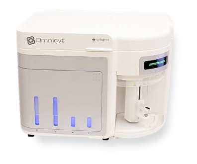
The Omnicyt™ CE-IVD flow cytometer provides a combination of precision, performance and reproducibility for your clinical cell analysis. In a field steadily evolving through standardization, Cytognos provides an important tool for the implementation of an efficient and reliable workflow, complementing the value of the Infinicyt™ software and the multiple reagent solutions which are based on the scientific excellence of EuroFlow™.
 |
 |
 |
 |
 |
 |
| Advantages | Features and specifications | Solutions | Software | Accesories | Support |
Advantages| Acoustic assisted hydrodynamic focusing system |
What’s acoustic focusing– High flow rates of highly diluted samples – Less doublets – Anti-clogging technology – Low fluid consumption: no fluidics cart is required |
|
| Automatic detection of bubbles |
The system is capable to detect bubbles stopping the acquisition and allowing to save previous data maintaining data files quality – No loss of valuable information – Save time |
|
| Possible to recover unused samples |
– No loss of sample – Safe acquisition |
|
| Different flow rates: 12,5, 25, 100, 200, 500 and 1.000 uL/min |
– High versatility – Quick acquisition at the fastest flow rate |
|
| Variable particle velocity: |
Variable particle velocity:
– Decreases acquisition time with highly diluted samples |
|
| Acquisition volume: 20 uL – 4 mL |
– High flexibility – Big accuracy |
|
| Data acquisition up to 35.000 events/second | – Allows flexibility and a quicker acquisition | |
| Flat top lasers |
What are flat top lasers?– Robust, sensitive and stable system – Less misalignment issues – Less need of calibration |
|
| Simmer mode: lasers switched on only during acquisition |
– Increases 10X lifetime – Less technical service issues |
|
| SSC detector for violet laser |
– Detection of small particles – Detection of erythrocytes simultaneously with any leucocyte population in no lyse no wash protocol with whole blood and bone marrow samples |
|
| No loss of information | – All the samples are saved even if they have not been recorded during acquisition | |
| No abort rate | – Possibility to save all events including electronic aborts | |
| Possible to record up to 20 million events with all parameters |
– Useful for the study of samples with rare populations – MRD studies |
Features and specifications| Optics | 3 lasers Blue laser: 488 nm, 50 mW Violet laser: 405 nm, 50 mW Red laser: 637 nm, 100 mW A fourth yellow laser (561 nm, 50 mW) enabling 16 parameters is available as an upgrade. 13 individual detectors: 11 fluorescent detectors 2 of light dispersion (FSC + SSC) Fluorescence sensitivity: <80 MESF for FITC <30 MESF for PE <70 MESF for APC Set of optimized filters for clinical cytometry most used fluorochromes: VL-1: 440/50 (PacB; BV421; SB436; PR436) VL-2: 512/25 (OC515; PacO*; AmCyan; BV510) VL-3: 603/48 (BV605; PacO*; SB600; PR600) VL-4: 710/50 (BV711; SB702) BL-1: 530/30 (FITC) BL-2: 574/26 (PE) BL-3: 695/40 (PerCP-Cy5.5) BL-4: 780/60 (PE-Cy7) RL-1: 670/14 (APC) RL-2: 720/30 (Alexa700; APC-A700) RL-3: 780/60 (APC-C750; APC-H7; APC-Cy7) *This fluorochrome can be detected by several filters. Flat-top parallel lasers with fixed alignment: High stability and sensitivity Lower calibration frequency |
|
| Fluidics | Volumetric system: Allows absolute cell counts (cells/µL) Low fluid consumption: no fluidics cart is required Allows different types of tubes (Ø 17×100 mm and 8.5×45 mm) Recovery of unacquired volumes in loaded samples Acoustic assisted hydrodynamic focusing: Higher reproducibility and accuracy even at high rates Flow rate between 12.5 and 1,000 µL/min: 12.5, 25, 100, 200, 500 and 1.000 µL/min Higher acquisition rate of diluted samples The acoustic focusing system works only from the flow rate: 100 µL/min. At 12.5 and 25 µL/min, the fluidic system works as a standard hydrodinamic system Anti-clogging technology: Reduced risk of clogging even with high concentration samples |
|
| Performance |
*This is the electronic resolution at optimal conditions; diagnostic samples are not intended to be acquired at this rate. |
|
| Acquisition software |
* Files may present higher percentage of doublets than expected.
|
|
| Quality |
|
|
| Cytometer dimensions | Benchtop cytometer with fluid bottles stored within:
No fluidics cart is required. |
|
| Consumables |
See the complete product list and more information |
Solutions
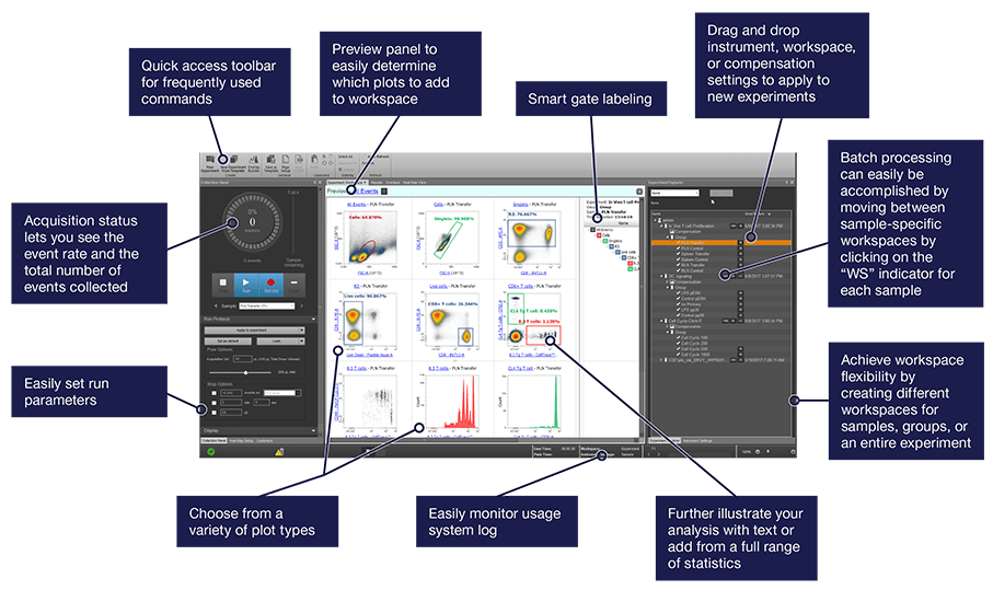
Software The Omnicyt software enables powerful user-defined experimental analysis via an intuitive, easy-to-use interface. Advanced tools provide simple acquisition and analysis for users at all experience levels.
Ease of use
Intuitive functionality-adjust flow rates, compensation and voltages through intuitive user interface
Less learning time-simple user interface with guided workflows
Easy instrument operation-automated maintenance and instrument performance monitoring
Speed
Quickly run flow cytometry experiments; less scrolling and clicking
Increase productivity; live-streaming update of statistics during acquisition of events
Fast refresh rates for large data sets of up to 20,000,000 events per sample and indeed, it’s posible to add more events with Append option
Guided functionality
Guided functions for automated or easy user-defined compensation
Automated maintenance: Start Up, Shutdown, Rinse, Santize Omnicyt™ SIP, Deep Clean, Sanitize, System Decontamination, Autosampler Calibration
Automated Data and Database backup to ensure data redundancy
© 2019 Thermo Fisher Scientific Inc. Used under permission. Life Technologies Corporation, a part of Thermo Fisher Scientific Inc. – www.thermofisher.com
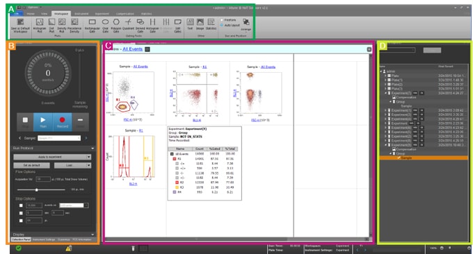
Visual management buttons and icons help navigate workflows and running experiments. The software allows for:
Customizable experimental workflows–pre-define settings for plots, gates, experiments, reports, and overlays
Preview screens–easy set-up by showing all plot combinations instantaneously for easy experimental set-up
Guided functions–for automated or easy user-defined compensation
Versatile
Heat map for easy setup of plate based assays
Sample Recovery to return unused sample to save precious samples
Hierarchy view of plots to instantly view complex gating strategies
Customizable
User customizable Wait-to-Record function
Ability to set user options for default settings for gates, plots, fonts, colors, and group/sample names
Visual appearance of the plots is completely customizable; fonts, colors, titles legends and much more can appear exactly the way you want
Publication quality data
Smartgate labeling to customize quad gate names and target names
Add text, statistics and even images to immediately present data
One-click saving of high resolution plots in a variety of file formats including XML, FCS, and even ZIP, for multiple files concurrently
Overlay module to perform comparative analysis of single and dual parameter data
Preview plots to instantly view all combinations of parameters in a file
User management
Levey-Jennings and Performance History reports of baseline and performance tests to monitor trends
Ability to create and manage multiple user accounts
System access based on user account privileges
System logs amount of time between user logon / logoff and number of tubes run per account per period of time
Note: The Omnicyt Flow Cytometer includes a dongle and 1 license for the Omnicyt Software plus a dongle and 1 license for the Infinicyt Software The Omnicyt software may be downloaded onto more than one computer, however the software will only run on computers that also have a dongle inserted. Users then can export FCS files in 3.0 and 3.1 format for use in other analysis programs such as Infinicyt Software.
© 2019 Thermo Fisher Scientific Inc. Used under permission. Life Technologies Corporation, a part of Thermo Fisher Scientific Inc. – www.thermofisher.com
Software simplifies data acquisition with a dashboard that provides the tools for immediately viewing acquisition status and for setting acquisition stopping criteria. The application settings feature automatically adjusts voltage settings to daily changes in cytometer performance, so that users can run existing applications more simply and quickly.
© 2019 Thermo Fisher Scientific Inc. Used under permission. Life Technologies Corporation, a part of Thermo Fisher Scientific Inc. – www.thermofisher.com
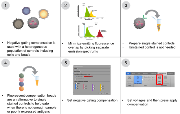
Compensation is both rapid and accurate using a guided software system. Users are only required to prepare the proper controls and adjust the voltage gates. The system adjusts the compensation and applies it to the samples.
Compensation tools
Automated compensation setup allows rapid and accurate compensation and helps to eliminate tedious trial and error adjustments
Both negative and unstained gating parameters are available
On-plot compensation for fine-tune adjustment
Modification of compensation to add or remove parameters as needed after compensation is set up
Setup and collect compensation controls directly from a plate
© 2019 Thermo Fisher Scientific Inc. Used under permission. Life Technologies Corporation, a part of Thermo Fisher Scientific Inc. – www.thermofisher.com
Accesories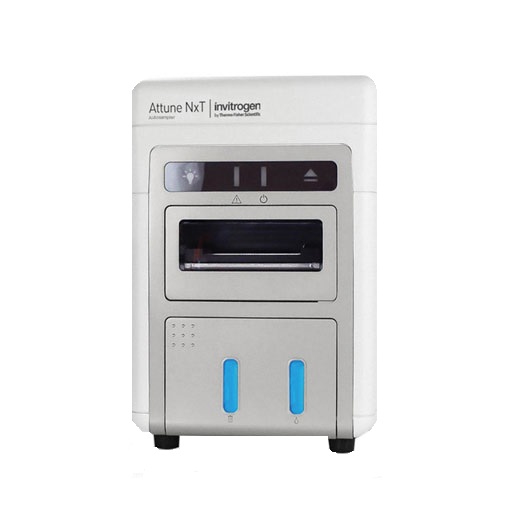 |
Autosampler
© 2019 Thermo Fisher Scientific Inc. Used under permission. Life Technologies Corporation, a part of Thermo Fisher Scientific Inc. – www.thermofisher.com |
|
Omnicyt™ No-Wash No-Lyse Filter Kit
|
 |
Omnicyt™ Yellow Laser
|